Using Thermography To Diagnose Horse Injuries
Without technology, veterinarians would have a much more challenging time locating and diagnosing problems with their non-verbal equine patients.
Time is another critical factor in the diagnosis and treatment of horse injuries and conditions, and because many animals display non-specific symptoms, they often have to endure lengthy delays before a definite diagnosis can be made. Finding answers can be frustrating and costly.
Enter infrared thermography which has proven to be a reliable and successful diagnostic and prognostic tool in horse practice, enabling veterinarians to diagnose and evaluate numerous disease conditions in horses in a shorter space of time as well provide information which assists in the follow-up treatment plan and recovery.
Conditions such as back pain, soft tissue injuries, arthritis and problems relating to flexor tendons, hooves and related structures are some of the issues where infrared thermography can play a positive role.
What is thermography?
Basically, thermography is a non-invasive heat-detecting technology that translates the surface temperature of an object into pictorial colour images. It has been used by the veterinary industry for several decades but in recent years, has come to the fore as a useful, cost-effective and reliable technology for preventative veterinary medicine, as well as diagnostics, treatment, prognosis and rehabilitation.
In the case of a horse, a thermal imaging camera is used to convert the infrared radiation that is emitted from the skin into electrical impulses that are then displayed on a colour screen. Depending on the severity of the inflammation and the associated increase in blood flow, the ‘hot spots’ are displayed on the videoscreen in red, orange or yellow (with red being the most severe).
A study involving 45 clinical cases where horses were examined by a thermal imager to see how useful the technology was for diagnosing vague diseased conditions found that the technology successfully located the area where the tissue has been injured and supplied veterinarians with important information regarding the likely time required for the horse to recover and the probability of full recovery.
In all the cases where a diagnosis was made (including tendonitis of flexors, summer sore and laminitis), thermal imaging resulted in a diagnosis being made up to two weeks before the appearance of clinical signs of the condition.
It’s a technology that can have significant benefits particularly when highly prized, valuable athletic animals are involved.
Take the case of four Arabian racehorses which had developed a restricted gait as a result of non-specific back pain. After the horses had been hospitalised, veterinarians made the clinical observation that because the animals’ longissimus muscle contracted involuntarily when the doctors applied pressure to the area, it indicated that there was a deeply sited non-identifiable lesion. The horses’ condition improved after contributory factors were removed (unfitted saddles over the area, heavyweight trainer atop the horse) and medications administered (including topical anti-inflammatories and intramuscular injections).
At the same time, thermal imagery of the horses prior to treatment showed increased skin temperature behind the withers and on both sides of the animals’ backs. This led to the diagnosis that the problem was inflammation of the longissimus muscles and related ligaments as well as severe inflammation of the spinals dorsi muscle.
Once treatment had been completed, there were no colour spots on the scans which indicated that the animals had recovered completely.
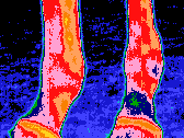
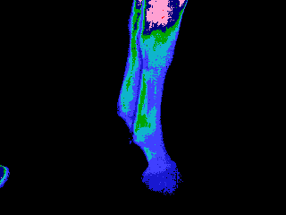
 |
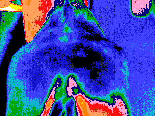 |
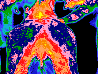
| Non-specific back pain, note the presence of yellow spot (primary lesion) and yellow /red-hot areas (Secondary lesion). | Same case after treatment, note the disappearance of the spot and the hot areas. |
That goes to show just why highly sensitive thermography can play such an important role in diagnosing horse injuries – notably pain caused by muscle injuries which are undetectable even by laboratory diagnosis.
Thermography not only can locate an area or areas of inflammation, but can also be used to highlight muscle atrophy before clinical symptoms appear – further underscoring its value and benefit as a diagnostic tool.
This early detection is vitally important in helping to prevent serious further injury. Researchers used thermography to look for areas of inflammation in horses that were showing a light degree of lameness – and results revealed coloured areas to indicate the problem areas. Treatment was successful but what was important was that the thermal imagery enabled veterinarians to pick up problem areas up to two weeks before the condition worsened into acute tendonitis – once again underscoring the effectiveness of thermography in avoiding further injury which may be serious.
 |
 |
 |
 |
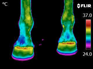
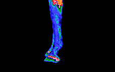
| Normal thermography of the flexor tendons |
Brown yellow spots over the inflamed superficial and deep flexors and suspensory ligaments | Red hot spots over the inflamed suspensory ligaments | Same case after treatment showing decreased thermal gradient |
An added advantage is that thermal imaging can be used to monitor the progress of a horse’s ongoing healing process – enabling better decision-making and planning and helping veterinarians do the most effective job they can in treating their equine patients.
Among the many other conditions in horses where thermography plays a valuable diagnostic role include navicular bursitis, pododermatitis, corns, thrush, tendonitis, summer sore and laminitis. Whilst it isn’t able to determine morphologic changes, it does locate the area of injured tissue and provide important information as to the horse’s recovery time and the probability of full recovery. Importantly however, the timing of the use of thermography in diagnostics is crucial. Early use (but after a thorough physical examination) will save time for the veterinarian, the horse’s owner and trainer as well as save money and prevent needless suffering for the horse.
As a leading veterinarian said: “Infrared thermography is a vital tool that helps us give animal owners good, solid, scientifically documented evidence on which we can base the best possible prognosis and plan for the animals’ recovery as well as accurately monitor the plan as we implement it.”
For qualified advice on infrared thermal imaging cameras or other specialised technical equipment, please contact us. We are a leading provider of technical equipment solutions to all industries across Australia and have a reputation for high quality, cost-effective and workable solutions to unique inspection challenges.
